
Contents
| Image | Title | Category | Type | Description | Updated |
|---|---|---|---|---|---|
|
slideToolkit | Software | Collection | SlideToolkit is a collection of command-line tools to assist with the automated histology analysis of whole-slide images. The publication linked in the "reference" details the actual workflow. This includes tools to organize the data, perform tiling and subsequent batch processing of the generated tiles in a cell profiler pipeline. All the tools are designed to run on a single PC or on a HPC system. The scripts in the toolkit are on github under MIT licence. |
05/03/2023 - 15:09 |
| |
Extract_Images | Software | Component | 05/20/2018 - 01:31 | |

|
Optical Flow | Software | Component | This protocol computes the optical flow of a 2D+T sequence. The results are displayed with flow arrows painted on top of the original sequence, and also with two additional sequences for the norm of the flow and a color-coded presentation of the flow following the Middlebury convention. |
05/15/2018 - 01:06 |
| |
LIF Projector | Software | Component | This macro is similar to the LIF_Extractor, but it will output Z-projection of each Z-stack. You can choose the type of projection in addition to the other options. Requires Bio-Formats plugin |
05/07/2018 - 16:47 |
|
|
Batch spot detection with custom output | Software | Workflow | Download the protocol,use and modify in Icy. It permits to detect spot with wavelet spot detector block. Input : loop on a folder Outputs: excel, binary, and detection screenshot |
10/30/2025 - 05:03 |
| |
LIF Extractor | Software | Component | This macro extracts .lei and .lif multichannel Z-stacks into multiple .tif stacks, splitting the channels into different stacks. Several options are possible such as background substraction, various filters, or optional reset of spatial scale. Requires Bio-Formats plugin |
05/07/2018 - 16:50 |
| |
OpenSlide | Software | Collection | >OpenSlide is a C library that provides a simple interface to read whole-slide images (also known as virtual slides). Python and Java bindings are also available. The Python binding includes a Deep Zoom generator and a simple web-based viewer. The Java binding includes a simple image viewer. |
12/07/2020 - 10:25 |
|
Pixel Classification using ilastik | Software | Component | This workflow classifies, or segments, the pixels of an image given user annotations. It is especially suited if the objects of interests are visually (brightness, color, texture) distinct from their surrounding. Users can iteratively select pixel features and provide pixel annotations through a live visualization of selected feature values and current prediction responses. Upon users' satisfaction, the workflow then predicts the remaining unprocessed image(s) regions or new images (as batch processing). |
10/18/2018 - 17:31 |

|
Counting foci in ImageJ | Software | Workflow | 06/09/2018 - 02:41 | |
|
|
3D estimation of synaptic vesicle distribution in serial section TEM (ssTEM) | Software | Workflow | An estimate of the shortest distance of vesicles to synaptic cleft is computed in 3D for serial section TEM. Unfortunately the the authors do not provide an implementation. Method: 1. Bias correction for inhomogene lighting 2. Image registration of TEM sections / stacks 3. Detection of vesicles & synaptic cleft (semi-automatic) 4. Compute distances in 3D | 09/12/2017 - 20:04 |
|
|
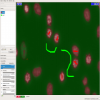
Ctrax - Caltech Multiple Fly Tracker | Software | Collection | Well maintained and documented project that includes a core tracking incl. GUI as well as Matlab toolboxes to (1) correct tracking results and (2) analyze fly behavior. >Ctrax is an open-source, freely available, machine vision program for estimating the positions and orientations of many walking flies, maintaining their individual identities over long periods of time. It was designed to allow high-throughput, quantitative analysis of behavior in freely moving flies. | 09/12/2017 - 20:04 |
|
|
Automated velocity mapping of migrating cell populations (AVeMap) | Software | Workflow | Requires Matlab Runtime Environment or Matlab. Source code (m-files) are downloaded. Software availability: AVeMap was developed under MATLAB (MathWorks). It is available as an executable, multiplatform program, together with source codes and documentation here, and the source code is also available as Supplementary Software. For practical reasons, this executable version, which does not require MATLAB, runs on a single processor. |
05/02/2023 - 19:04 |
|
|
Uneven illumination correction | Software | Workflow | Illumination correction is often important for both accurate segmentation and for intensity measurements. This example shows how the CorrectIlluminationCalculate and CorrectIlluminationApply modules are used to compensate for the non-uniformities in illumination often present in microscopy images. |
01/31/2018 - 16:37 |
|
Wound healing assay: analysis in CellProfiler | Software | Workflow |
Download package also contains example images. |
03/10/2023 - 23:18 |

|
CellProfiler Comet assay/DNA damage assay | Software | Workflow | quote
Example Images: Packaged together with the cellprofiler pipeline file. |
05/16/2018 - 21:56 |

|
Leaf Infection Tools | Software | Collection | The Leaf Infection Tools allow to measure the area of leaves, of two stainings in different channels and of the overlap region of the two stainings. See: http://dev.mri.cnrs.fr/projects/imagej-macros/wiki/Leaf_Infection_Tools Test image: http://biii.eu/node/1143 |
11/16/2018 - 09:46 |
|
Oufti | Software | Collection | 05/26/2018 - 00:52 | |
|
SparkMaster | Software | Workflow | Analyzing Ca2+ sparks ImageJ plugin to detect and measure Ca2+ sparks in linescan images, described in Picht et. al. (2007). The algorithm is based on that described by Cheng et al. (1999). Care should be taken to ensure that detections belong to 'true' events, as without any additional background subtraction steps the algorithm is not appropriate for images in which the baseline fluorescence varies substantially. |
05/20/2018 - 23:07 |

|
Object Classification using ilastik | Software | Component | This workflow classifies objects based on object-level features (e.g. intensity based, morphology based, etc) and user annotations. It needs segmentation images besides the raw image data. Segmentation images can be obtained from ilastik pixel classification, or binary segmentation images from other tools. Within the object classification, one can prefilter objects through thresholds (on pixel probability image) or object sizes (on segmentation image). Outputs are predicted classification label images. Selected features can also be exported. |
05/16/2018 - 02:00 |
|
|
Automatic 2D/3D Tracking using ilastik | Software | Workflow | This workflow is used to track multiple (appear/disappear, dividing and merging) objects in presumably big 2D+t or 3D+t datasets. It is best suitable for roundish objects or spots. Tracking is done through segmentation, which can be obtained from ilastik pixel classification, or imported from other tools. Users should provide a few object level labels, and the software predicts results on the rest of the image or new images with similar image characteristics. |
04/28/2023 - 15:22 |