
Contents
| Image | Title | Category | Type | Description | Updated |
|---|---|---|---|---|---|

|
Measuring confluence in adherent cell cultures | Software | Workflow | This publication describes a very simple protocol to acquire images of adherent cell cultures over time and how to process these images in ImageJ to measure the area fraction (confluence). | 09/12/2017 - 20:04 |
|
|
Noise Suppression using Simple Spatial Filters | Software | Component | Simple spatial filters can be used to suppress noise in raw image data (i.e. by averaging intensities). The best choice of filter depends on the nature of the noise, but Gaussian filtering works well for Poisson noise (i.e. commonly observed photon-counting shot noise); whereas a median filter is ideal for salt-and-pepper noise. A larger filter radius leads to stronger noise suppression but more blurring. The URL above describes the simple 2D spatial filters available in ImageJ, but similar filters are available in most software. |
11/26/2018 - 09:32 |
|
|
Interactive Density Counting using ilastik | Software | Workflow | This workflow estimates (densely distributed) object counts by the density of objects in the image without performing segmentation or object detection. Current version only works for 2D images of roundish objects with similar sizes on relatively homogeneous background. Users should provide a few labels of background and objects (especially on clustered objects), and the tool predicts the density of objects on the entire image. Counting is then estimated by integrating the density values on the whole image or specified rectangular regions of interests. | 09/13/2017 - 12:14 |

|
Artemia color analysis | Software | Workflow | The Artemia Tools help to calculate the normalized redness of Artemia in color images. See: http://dev.mri.cnrs.fr/projects/imagej-macros/wiki/Artemia_Tools Test images: http://biii.eu/node/1139 |
09/13/2017 - 16:02 |
|
VSI File Extractor (ImageJ) | Software | Component | This tool allows for extraction of image series from Olympus Slide Scanners. These VSI files usually contain several images that are too big to load into memory (>50k x 50k pixels). It was written and tested on Fiji and is available from a Fiji Update Site: http://fiji.sc/List_of_update_sites |
03/02/2020 - 20:56 |
|
|
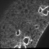
Human cytoplasm-nucleus translocation assay using CellProfiler | Software | Workflow | In this human cytoplasm-nucleus translocation assay, learn how to load a previously calculated illumination correction function for two separate channels, measure protein content in the nucleus and cytoplasm, and calculate the ratio as a measure of translocation. This is a clumpy cell type, so studying the settings in primary object identification may be helpful for users interested in the more advanced options that module offers. More about these images can be found at the BBBC. |
03/03/2020 - 20:45 |
|
|
Imaris Tracking | Software | Workflow | ImarisTrack allows 3D tracking of spots and objects, with straightforward manual adjustment of automatic tracking results. | 09/13/2017 - 12:14 |
|
|
Measure distance of organelles from nucleus in 3D | Software | Workflow | 06/09/2018 - 02:08 | |

|
Find cells using nuclear and membrane fluorescence | Software | Workflow |
|
04/29/2023 - 15:29 |
|
|
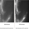
White Balance | Software | Component | The Macro processes a composite picture in ImageJ/Fiji and outputs a color-balanced merged RGB image. To calculate the white balance, a rectangle at coordinates (x=100, y=100) and of size (w=100 pixels, h=100 pixels) is used. These values can be changed to make sure that a background region is taken for the calculation in the line: makeRectangle(100,100,100,100). The user could be prompted to draw the region by removing the signs // in the line: // waitForUser("Please draw a region in the background"); |
05/21/2018 - 01:37 |
|
|
Cells tracking in tissue | Software | Workflow | Plot the centroid tracks and area evolution of the cells of a tissue with membrane labelling. | 09/13/2017 - 12:14 |

|
2D and 3D mitochondrial shape analysis and network properties | Software | Workflow | The original paper describes a method to analyze mitochondrial morphology in 2D and 3D. |
04/26/2023 - 15:43 |
| |
Object Tracking and Metadata Management | Software | Workflow | 05/16/2018 - 02:11 | |
|
Image denoising using Matlab | Software | Component | If your images are corrupted by a strong dominant Gaussian noise you can try this simple filter. It is based on thresholding in the DCT domain and is usually vastly superior to typical Gaussian filtering in term of detail preservation / noise reduction trade-off. |
04/28/2023 - 16:56 |

|
Interactive Volume of Interest Extractor in Large 3D Stacks | Software | Component | This macro allows to interact with a large, single channel, z-stack (possibly exceeding the main memory of the computer) and to extract a volume of interest by marking several reference points. |
04/28/2023 - 16:48 |
|
|
Microtubule Length Analysis | Software | Workflow | Task Quantify the length of microtubules (MT) and the MT average density per cell. Workflow descriptions Simple two step workflow, allowing visual & manual correction of microtubule between the 2 steps. Batch measurement of microtubule lengths for multiple images is achieved by segmenting the MTs and then their skeletonizations. The number of pixels in the microtubule is proportional to their length, so the length can be estimated. Script Workflow is written as an ImageJ macro (Fiji) with following steps: |
05/03/2023 - 10:39 |
| |
(Z,T,C) Stitcher | Software | Component | This macro can stitch a (Z,T,C) data set with virtually no limit on the number of Z slices and time frames. The input to the macro is a folder with the raw tiff images (one image per file) as typically exported by motorized microscopes. These files must all be stores in the same folder and the file naming should ideally comply to OME-TIFF. The macro is however quite flexible: Only --X, --Y and --Z fields with user defined number of digits are compulsory. --T, --C and --L fields with user defined number of digits are necessary for multiple time frames / channels data sets. |
04/28/2023 - 16:40 |
|
(Z,T) Hyperstack Stitcher | Software | Component | This macro builds a stitched image from a muti-position 3D + time hyperstack. The XY positions of the montage should be coded as channels in the input hyperstack. Channel ordering can be configured in the dialog box to adapt to Column/Row and Meander/Comb configurations: The images should appear in this order when browsing the hyperstack with the channel slider. Fine stitching is supported (requires sufficient overlap between the views). |
04/28/2023 - 16:32 |
|
|
Marker-controlled Watershed (ImageJ) | Software | Component | Marker-controlled Watershed is an ImageJ/Fiji plugin to segment grayscale images of any type (8, 16 and 32-bit) in 2D and 3D based on the marker-controlled watershed algorithm (Meyer and Beucher, 1990). This algorithm considers the input image as a topographic surface (where higher pixel values mean higher altitude) and simulates its flooding from specific seed points or markers. |
03/02/2020 - 21:01 |

|
ImmunoRatio | Software | Workflow | Analyzing ER, PR, and Ki-67 immunohistochemistry ImmunoRatio is an ImageJ plugin to quantify haematoxylin and DAB-stained tissue sections by measuring the percentage of positively stained nuclear area (labeling index), described in [bib]2452[/bib]. Notes for use: |
04/28/2018 - 23:13 |