
Contents
| Image | Title | Category | Type | Description | Updated |
|---|---|---|---|---|---|

|
Neuron Tracing Vaa3D (MOST) | Software | Workflow | 3D Neuron Tracing with a Dockerized version of Vaa3D MOST Raytracer. |
05/01/2023 - 18:27 |
|
|
Neuron Tracing Vaa3D (MST) | Software | Workflow | 3D Neuron Tracing using Dockerized version of Vaa3D Minimum Spanning Tree (MST). |
05/17/2023 - 18:53 |
|
|
Neuron Tracing 3D (Rivuletpy) | Software | Workflow | Rivuletpy dockerised workflow for BIAFLOWS. |
05/17/2023 - 20:30 |
|
|
Neuron Tracing Vaa3D (App2) | Software | Workflow | Vaa3d All-Path-Pruning 2.0 (APP2) dockerised workflow for BIAFLOWS. |
05/17/2023 - 19:43 |
|
|
Object Tracking (MU-Lux-CZ) | Software | Workflow | Cell tracking using MU-Lux-CZ algorithm. Dockerized Workflow for BIAFLOWS implemented by Martin Maska (Masaryk University). |
05/01/2023 - 18:43 |
|
|
Object Tracking (Octave) | Software | Workflow | Nuclei tracking in 2D time-lapse with Octave tracker (adapted from Matlab LOBSTER version). |
05/17/2023 - 19:41 |
|
|
Object Tracking (ImageJ) | Software | Workflow | Object tracking. For each time-frame, an image mask is obtained from median filtering (user defined radius), thresholding (user defined level) and hole filling. Convex objects are split apart by distance map watershed from regional intensity maxima (user defined noise tolerance), eroded (user defined radius) and analyzed as 3D particles (assuming some overlap between objects from a frame to the next frame). Finally, division events are analyzed and accounted for to relabel objects. |
05/17/2023 - 19:40 |
|
|
pyimagej | Software | Collection | pyimagej provides a set of wrapper functions for integration between ImageJ and Python. It also provides a high-level entry point for invoking ImageJ server APIs. |
04/29/2023 - 10:42 |
|
|
Nuclei Tracking (TrackMate) | Software | Workflow | Track non-dividing particles in 2D time-lapse image. |
05/17/2023 - 20:22 |
|
|
Particle Tracking (ImageJ) | Software | Workflow | Particle tracking in 2D time-lapse based on linking closest regional intensity minima (user defined noise tolerance) detected from Laplacian of Gaussian filtered images (user defined radius). A maximum linking distance is set (user defined). |
05/17/2023 - 20:23 |
|
|
Nuclei Segmentation 3D (NucleiSegmentation3D-ilastik) | Software | Workflow | Execute Nuclei Segmentation in 3D images using pixel classification with ilastik. |
04/28/2023 - 13:26 |
|
|
Nuclei Segmentation 2D watershed (NucSeg3DThr-ImageJ) | Software | Workflow | The macro will segment nuclei and separate clustered nuclei in a 3D image using a 2D Gaussian blur, followed by Thresholding, 2D hole filling and a 2D watershed. As a result an index-mask image is written for each input image. |
05/17/2023 - 19:46 |
|
|
Nuclei Segmentation (U-Net) | Software | Workflow | U-Net segmentation as presented in Reference Publication. The model predicts three classes: background, edge and foreground. The model was trained with Kaggle Data Science Bowl (DSB) 2018 training set. |
05/17/2023 - 19:48 |
|
|
Nuclei Segmentation (DeepCell) | Software | Workflow | Nuclei Segmentation using Deep Learning for individual cell analysis (DeepCell). |
05/17/2023 - 19:51 |
|
|
FeatureJ Hessian | Software | Component | This plugin computes for each image element (pixel/voxel) the eigenvalues of the Hessian, which can be used for example to discriminate locally between plate-like, line-like, and blob-like image structures |
05/17/2023 - 20:19 |
|
|
Spot Detection 3D Hessian (ImageJ) | Software | Workflow | 3D spot detection using the Determinant of Hessian (DoH) and the detection of 3D minima. |
05/17/2023 - 20:25 |
|
|
Spot Detection 3D (Icy) | Software | Workflow | Spot detection in 3D images by Wavelet Adaptive Threshold in Icy. |
05/24/2023 - 16:31 |
|
|
Spot Detection Dmap (ImageJ) | Software | Workflow | This workflow detects spots in a 2D image by filtering the image by Laplacian of Gaussian (user defined radius), thresholding (user defined threshold) and finding local intensity maxima in mask distance map (Dmap). |
05/24/2023 - 16:35 |
|
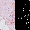
Nuclei segmentation in histopathology images | Dataset | This dataset has been announced in the paper "Segmentation of Nuclei in Histopathology Images by deep regression of the distance map" in Transaction on Medical Imaging 2019. |
10/27/2025 - 09:13 | |

|
MoNuSeg - Multi-organ nuclei segmentation challenge | Dataset | The dataset for this challenge was obtained by carefully annotating tissue images of several patients with tumors of different organs and who were diagnosed at multiple hospitals. This dataset was created by downloading H&E stained tissue images captured at 40x magnification from TCGA archive. H&E staining is a routine protocol to enhance the contrast of a tissue section and is commonly used for tumor assessment (grading, staging, etc.). |
10/28/2025 - 01:51 |