
- 188 views
Fluorescence microscopy cannot be used to image human embryos to determine embryo viability for in vitro fertilization because the introduction of exogenous fluorescent dyes is considered a toxic procedure. As a result, embryo viability has been measured primarily using differential interference contrast (DIC). A human can readily segment the embryo (and, to some extent, individual cells) in a DIC image, but automatic segmentation remains a challenge due to the cosine-dependent shading inherent in DIC images.
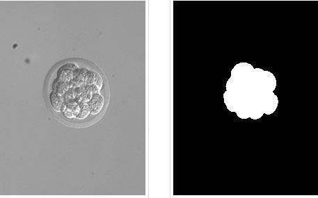
For the purpose of collecting ground truth, the samples were Hoechst-stained and imaged by confocal microscopy, and the cells were counted by a simple human. A tab-delimited text file contains cell counts in each of the 15 images.
There are 15 images. The images were acquired using a Nikon Eclipse TE200 microscope with a 20x, 0.45 NA objective lens and a 0.52 NA condenser lens, and are provided courtesy of the W.M. Keck 3D Fusion Microscope Facility at Northeastern University. Each image contains 640 x 480 pixels with an approximate size of 0.42 x 0.42 μm.