
Contents
| Image | Title | Category | Type | Description | Updated |
|---|---|---|---|---|---|

|
arivis Vision4D | Software | Collection | arivis Vision4D is a modular software for working with multi-channel 2D, 3D and 4D images of almost unlimited size independent of available RAM. Many imaging systems, such as high speed confocal, Light Sheet/ SPIM and 2 Photon systems, can produce a huge amount of multi-channel data, which arivis Vision4D handles without constraints. |
03/05/2020 - 13:11 |
|
|
3D segmentation (reconstruction) and modeling using Free-D | Software | Workflow | Free-D (http://free-d.versailles.inra.fr/) is a 3D reconstruction and modeling software. It is multiplatform, free (but not open source) tool for academic research and teaching. Here is how to proceed, using Free-D: 1. Segmentation: * load (a collection of) individual 3d stacks * (optional for serial sections) perform a 2D registration to align image slices * segment/reconstruct 3D contours using snakes * segment 3D spots 2. Construct average cell: |
04/29/2023 - 13:43 |
|
|
ThunderSTORM | Software | Collection | ThunderSTORM is an open-source, interactive, and modular plug-in for ImageJ designed for automated processing, analysis, and visualization of data acquired by single molecule localization microscopy methods such as PALM and STORM. Our philosophy in developing ThunderSTORM has been to offer an extensive collection of processing and post-processing methods so that users can easily adapt the process of analysis to their data. |
03/05/2020 - 16:36 |
|
|
MicrobeJ | Software | Collection | This is an ImageJ plugin to analyze bacterial cells. It provides a user-friendly interface and a powerful suite of detection, analysis and data presentation tools. It works with individual phase or fluorescence images as well as stacks, hyperstacks, and folders of any of these types. Even large image sets are analyzed rapidly generating raw tabular data that can either be saved or copied as is, or have additional statistical analysis performed and graphically represented directly from within MicrobeJ, making it an all-in-one image analysis solution. |
04/26/2023 - 13:14 |
|
|
MultiStackReg | Software | Component | This ImageJ plugin aligns the slices of a stack just like the stackreg plugin on which it is built. It allows to save the transformations and to apply them to another stack. It furthermore allows to register two stacks. |
04/26/2023 - 13:35 |

|
classification of hemp fibers based on morphological features | Software | Workflow |
In this workflow, you can use MorphoLibJ to generate accurate morphometric measurements |
08/16/2018 - 17:37 |
|
SIMToolbox | Software | Collection | SIMToolbox: a MATLAB toolbox for structured illumination microscopy SIMToolbox is an open-source, modular set of functions for MATLAB designed for processing data acquired by structured illumination microscopy. Both optical sectioning and super-resolution applications are supported. The software is also capable of maximum a posteriori probability image estimation (MAP-SIM), an alternative method for reconstruction of structured illumination images. |
11/26/2018 - 09:30 |
|
fairSIM | Software | Collection | An easy-to-use plugin that provides SR-SIM reconstructions for a wide range of SR-SIM platforms directly within ImageJ. For research groups developing their own implementations of super-resolution structured illumination microscopy, fairSIM takes away the hurdle of generating yet another implementation of the reconstruction algorithm. For users of commercial microscopes, it offers an additional, in-depth analysis option for their data independent of specific operating systems. |
01/11/2019 - 12:14 |

|
Grayscale granulometry | Software | Component | This imageJ/Fiji plugin provides an analysis of the granulometry inside an image by mathematical morphology. It has sevral option for the structuring element to be used, and the size domain to be tested. The output will be both a curve of the remaining content of the image against the growing size of the structuring element, and the corresponding results table that could be then exported. It can deal with grayscale images directly, no need to segment the image first. |
10/18/2018 - 17:35 |

|
KNOSSOS - 3D image visualization and annotation tool | Software | Collection | It is a tool to visualize and annotate volume image data of electron microscopy. Users can annotate objects (e.g. neurons) and skeleton structures. It provides the ability to overlaying the image data with user annotations, representing the spatial structure and the connectivity of labeled objects, and displaying a three dimensional model of it. It can be extended by plugins written in python. A similar, web-based implementation is being developed at webknossos.info. Example datasets are also available. |
04/26/2023 - 13:23 |

|
MorphoLibJ | Software | Collection | MorphoLibJ is a library of plugin for ImageJ with functionalities for image processing such as filtering, reconstructing, segmenting, etc... Tools are based on Mathematical morphology with more rigorous mathematical approach than in the standard tools of ImageJ in particular for surface (or perimeter) measurements which are usually based on voxel counting. http://imagej.net/MorphoLibJ#Measurements Among the features: |
04/25/2023 - 19:17 |
|
|
Fourier Ring Correlation | Software | Component | Calculate the Fourier ring correlation (FRC). The FRC can be used as a resolution criterion for super resolution microscopy. The Plugin can display a plot of the FRC curve, along with the LOESS smoothed version of the curve. Finally it displays the threshold method used and the intersection of the FRC with the threshold, providing the FIRE number. It can be used on two open images or on pairs of images in batch mode. 2654 2655 |
10/21/2019 - 10:44 |
|
|
MorphoLibJ: using morphological reconstructions to isolate objects | Software | Workflow | When trying to isolate objects, one strategy might be to use regular morphological operations (opening/closing) to remove small objects that are not of interest. In case small objects are made of a large number of pixels, this operation might impair the remaining objects' contours. An alternative strategy might be to use morphological reconstruction. In short, seed is placed on the image, on objects, then conditional dilation is performed from those seeds. Here is how to proceed, using MorphoLibJ: |
05/03/2023 - 16:19 |

|
neuTube (or NeuTu) | Software | Collection, Component | neuTube is a collection of neuron reconstruction tools from fluorescence microscope images. It has an interactive system with a 3D viewer, which can be clicked in 3D and perform neuron tracing automatically and semi-automatically. It can automatically recognize branching points as junctions. Traced neurons can be exported to swc format, which could be imported by various software packages. neuTube has Win and Mac OS standalone executable builds and may also be installed by manual compilation. In addition, neuTube can be used as a plugin in Vaa3D.
|
04/27/2023 - 13:30 |
|
|
BiDiFuse | Software | Component | to be completed |
10/21/2019 - 10:48 |
|
|
iterative closest point registration | Software | Component | The ICP algorithm takes two point clouds as an input and return the rigid transformation (rotation matrix R and translation vector T), that best aligns the point clouds. Example: [R,T] = icp(q,p,10); Aligns the points of p to the points q with 10 iterations of the algorithm. The transformation is then applied using R*p + repmat(T,1,length(p)); The file has implemented both point to point and point to plane as well as a couple of other features such as extrapolation, weighting functions, edge point rejection, etc. |
04/26/2023 - 09:49 |
|
|
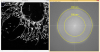
Hough Circles | Software | Component | This plugin applies the Hough Transform for Circles to an 8-Bit image, shows the resulting Hough Space in a new window and marks the centers of the found circles. |
10/21/2019 - 10:59 |
|
|
ABSnake | Software | Component | ABSnake can segment complex structures in 2D images as well as 3D or temporal images. It uses a new active contour model based on a geometrical approach for correctly following invaginated structures. |
05/03/2023 - 17:11 |

|
Lama: The LocAlization Microscopy Analyzer | Software | Collection | LocAlization Microscopy Analyzer (LAMA) is a software tool that contains several well-established data post processing algorithms for single-molecule localization microscopy (SMLM) data. LAMA has implemented algorithms for cluster analysis, colocalization analysis, localization precision estimation and image registration. LAMA works with a graphical user interface (GUI), and accepts simple input data formats as supported by various single- molecule localization software tools. |
05/03/2023 - 15:27 |
|
|
MosaicIA | Software | Component | MosaicIA is a tool to analyze the spatial distribution of objects in images. It estimates from an observed particle or object distribution what hypothetical interaction between the objects is most likely to have created this distribution. |
10/21/2019 - 11:06 |