
Contents
| Image | Title | Category | Type | Description | Updated |
|---|---|---|---|---|---|
|
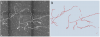
TReMAP | Software | Component | "we present a new fully automated 3D reconstruction algorithm, called TReMAP, short for Tracing, Reverse Mapping and Assembling of 2D Projections. Instead of tracing a 3D image directly in the 3D space as seen in majority of the tracing methods, we first trace the 2D projection trees in 2Dplanes, followed by reverse-mapping the resulting 2D tracing results back into the 3D space as 3D curves; then we use a minimal spanning tree (MST) method to assemble all the 3D curves to generate the final 3D reconstruction. |
04/11/2018 - 12:38 |

|
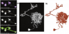
APP2 | Software | Component | All-path-pruning 2.0 (APP2) is a component of Vaa3D. APP2 prunes an initial reconstruction tree of a neuron’s morphology using a long-segment-first hierarchical procedure instead of the original termini-first-search process in APP. APP2 computes the distance transform of all image voxels directly for a gray-scale image, without the need to binarize the image before invoking the conventional distance transform. APP2 uses a fast-marching algorithm-based method to compute the initial reconstruction trees without pre-computing a large graph. This method allows to trace large images. |
10/16/2018 - 15:07 |
|
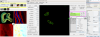
Vaa3D | Software | Collection | Vaa3D is a handy, fast, and versatile 3D/4D/5D Image Visualization and Analysis System for Bioimages and Surface Objects. It also provides many unique functions that you may not find in other software. It is Open Source, and supports a very simple and powerful plugin interface and thus can be extended and enhanced easily. |
05/03/2023 - 16:52 |
|
APP (All-path pruning) | Software | Component | "We have developed an automatic graph algorithm, called the all-path pruning (APP), to trace the 3D structure of a neuron. To avoid potential mis-tracing of some parts of a neuron, an APP first produces an initial over-reconstruction, by tracing the optimal geodesic shortest path from the seed location to every possible destination voxel/pixel location in the image. |
04/11/2018 - 12:51 |
|
QuimP | Software | Collection | Summary QuimP is software for tracking cellular shape changes and dynamic distributions of fluorescent reporters at the cell membrane. QuimP's unique selling point is the possibility to aggregate data from many cells in form of spatio-temporal maps of dynamic events, independently of cell size and shape. QuimP has been successfully applied to address a wide range of problems related to cell movement in many different cell types. Introduction |
05/02/2023 - 12:39 |
|
Scipion | Software | Collection | Scipion is an image processing framework for obtaining 3D models of macromolecular complexes using Electron Microscopy (3DEM). It integrates several software packages and presents a unified interface for both biologists and developers. Scipion allows you to execute workflows combining different software tools, while taking care of formats and conversions. Additionally, all steps are tracked and can be reproduced later on. |
10/13/2017 - 16:59 |
| Understanding the fundamental mechanisms of biofilms development and dispersal: BIAM (Biofilm Intensity and Architecture Measurement), a new tool for studying biofilms as a function of their architecture and fluorescence intensity | Training Material | Confocal laser scanning microscopy (CLSM) is one of the most relevant technologies for studying biofilms in situ. Several tools have been developed to investigate and quantify the architecture of biofilms. However, an approach to quantify correctly the evolution of intensity of a fluorescent signal as a function of the structural parameters of a live biofilm is still lacking. Here we present a tool developed in the ImageJ open source software that can be used to extract both structural and fluorescence intensity from CLSM data: BIAM (Biofilm Intensity and Architecture Measurement). |
01/28/2018 - 12:53 | ||
|
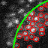
Angiogenesis / Sprout Analyzer (ImageJ) | Software | Component | The Sprout Morphology plugin measures sprout number, length, width and cell density of endothelial cell (EC) sprouts grown in a bead sprouting assay. It optionally includes measuring the coverage of these sprouts with pericytes included in the assay, as well as the endothelial cell/pericyte ratio. |
04/27/2023 - 16:56 |
|
SOAX | Software | Collection | SOAX is an open source software tool to extract the centerlines, junctions and filament lengths of biopolymer networks in 2D and 3D images. It facilitates quantitative, reproducible and objective analysis of the image data. The underlying method of SOAX uses multiple Stretching Open Active Contours (SOACs) that are automatically initialized at image intensity ridges and then stretch along the centerlines of filaments in the network. SOACs can merge, stop at junctions, and reconfigure with others to allow smooth crossing at junctions of filaments. |
05/03/2023 - 10:52 |
|
performing automatic registration for CLEM | Software | Workflow | This is an example workflow of how to perform automatic registration by - first detecting spots in both images using wavelet segmentation (with different scale according to the image scale) - second using Ec-Clem autofinder to register both images Click on a block to know more about a tool. Non referenced tools are non clickable. |
03/01/2020 - 11:31 |
|
Spot Detector (Icy) | Software | Component | Spot detector detects and counts spots, based on wavelet transform. |
03/03/2020 - 14:37 |
|
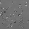
2D bright field yeast cell images with ground truth annotations | Dataset | Dataset used to evaluate the method described in "Yeast cell detection and segmentation in bright field microscopy", ISBI 2014 (DOI: 10.1109/ISBI.2014.6868107). |
02/05/2019 - 12:18 | |
|
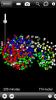
WormGUIDES Atlas | Software | Collection | WormGUIDES Atlas is an interactive 4D portrayal of neural development in C. elegans. It will ultimately contain nuclear positions for every cell in the embryo, identified and tracked from the 2 cell stage until hatching. Single-cell and subcellular information, including neural outgrowth dynamics for each cell as well as cell function, gene expression, the adult neural connectome and related literature will be collated for each cell from public sources and also integrated with the atlas model. WormGUIDES Atlas integrates tools for exploratory data analyses and insight sharing. |
04/30/2023 - 18:33 |

|
JACoP | Software | Component | This ImageJ plug-in is a compilation of co-localization tools. It allows: -Calculating a set of commonly used co-localization indicators: Pearson's coefficient Overlap coefficient k1 & k2 coefficients Manders' coefficient Generating commonly used visualizations: -Cytofluorogram Having access to more recently published methods: -Costes' automatic threshold Li's ICA Costes' randomization Objects based methods (2 methods: distances between centres and centre-particle coincidence) |
11/16/2018 - 09:47 |

|
ImageJ | Software | Collection | Bio Image Analysis tool from REF |
09/13/2017 - 12:12 |

|
Image Composite Editor | Software | Collection | ICE (Image Composite Editor) is a fast, fully automatic software by Microsoft that can create large montages from overlapping images. Although it is tailored around the task of stitching together images from a photo camera, it also works on biological images taken from light and electron microscopes. |
09/13/2017 - 12:16 |

|
GelBandFitter | Software | Collection | GelBandFitter is a user-friendly software specific for analysis of protein gels and estimation of relative protein content. Using non-linear regression methods to fit mathematical functions to densitometry profiles, it is able to estimate content from protein bands that partially overlap. The software is available either as Matlab code (Optimization toolbox required) or a Windows executable. Reference: Mitov, M. I., Greaser, M. L., & Campbell, K. S. (2009). GelBandFitter A computer program for analysis of closely spaced electrophoretic and immunoblotted bands. |
10/18/2018 - 17:35 |
|
Super-Resolution Radial Fluctuations (SRRF) | Software | Component | SRRF is a high-performance analytical approach for Live-cell Super-Resolution Microscopy, provided as a fast GPU-enabled ImageJ plugin. SRRF is capable of extracting high-fidelity super-resolution information from TIRF, widefield and confocals using conventional fluorophores such as GFP. SRRF is capable of live-cell imaging over timescales ranging from minutes to hours. |
05/03/2023 - 11:13 |
|
Spotsizer | Software | Collection | Spotsizer is a software tool that automates analysis of large volumes of photographic images of growing microbes. |
03/03/2020 - 17:46 |

|
CellProfiler | Software | Collection | CellProfiler is free, open-source software for quantitative analysis of biological images. |
10/17/2019 - 16:24 |