
Contents
| Image | Title | Category | Type | Description | Updated |
|---|---|---|---|---|---|

|
ATLAS Vesicle segmentation method | Software | Component | Part of ATLAS software Comment / Instructions: You can upload your image at the Mobyle@SERPICO portal and download the result. The workflow is only available online, i.e. no download possible. |
05/27/2018 - 18:58 |

|
Arabidopsis plants (low resolution) | Dataset | 09/13/2017 - 18:15 | ||

|
Arabidopsis plants (high resolution) | Dataset | 09/13/2017 - 18:12 | ||

|
FFmpeg | Software | Collection | 03/11/2019 - 17:34 | |

|
leaves stained with gfp and rfp | Dataset | 09/13/2017 - 17:30 | ||

|
GDSC plugins | Software | Collection | 12/16/2018 - 16:47 | |

|
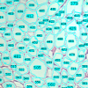
Artemia color images | Dataset | 09/13/2017 - 16:00 | ||
|
Tubeness | Software | Component | This plugin filters a 3D image stack (or 2D image) to produce a score for how "tube-like" each point in the image is. This is useful as a preprocessing step for tracing neurons or blood vessels, for example. For 3D image stacks, the plugin uses the eigenvalues of the Hessian matrix to calculate this measure of "tubeness", using a metrics mentioned in Sato et al 1997 ¹: if the larger two eigenvalues (λ₂ and λ₃) are both negative then value is √(λ₂λ₃), otherwise the value is 0. For 2D images, if the large eigenvalue is negative, we return its absolute value and otherwise return 0. |
10/21/2019 - 11:10 |

|
Microtubules 3D | Dataset | 05/03/2023 - 10:48 | ||
|
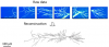
Reconstruct | Software | Collection | By combining multiple image alignment and tracing into one program, Reconstruct (TM) allows images to be processed more efficiently. Tracing can be done directly on the transformed images and alignments can be asily modified. Reconstruct (TM) was developed from years of experience working with high magnification serial section images of brain tissue. (Extracted from User Manual) |
09/13/2017 - 14:29 |
|
Adipocyte quantification ImageJ by Baecker | Software | Workflow | The Adipocytes Tools help to analyze fat cells in images from histological section. This is a rather general cell segmentation approach. It can be adapted to different situations via the parameters. This means that you have to find the right parameters for your application. Sample Image: [0178_x5_3.tif](http://dev.mri.cnrs.fr/attachments/190/0178_x5_3.tif) |
05/21/2018 - 00:38 |

|
Adipocyte quantification MATLAB | Software | Workflow | 05/21/2018 - 00:34 | |

|
JFilament | Software | Component | JFilament is an ImageJ plugin for segmentation and tracking of 2D and 3D filaments in fluorescenece microscopy images. The main algorithm used in Jfilament is "Stretching Open Active Contours" (SOAC). In order to use this method, the user must define seed points in the image where the SOAC method will begin. JFilament also includes 2D "closed" active contours which can be used for tasks such as segmentation and tracking of cell boundaries.
|
09/13/2017 - 13:38 |

|
Directionality | Software | Component | This plugin is used to infer the preferred orientation of structures present in the input image. It computes a histogram indicating the amount of structures in a given direction. Images with completely isotropic content are expected to give a flat histogram, whereas images in which there is a preferred orientation are expected to give a histogram with a peak at that orientation. On top of the histogram, the plugin tries to generate statistics on the highest peak found. |
10/30/2025 - 05:10 |

|
Arabidopsis thaliana seedlings | Dataset | Arabidopsis thaliana seedlings in liquid culture |
09/13/2017 - 12:16 | |

|
KNIME | Software | 09/13/2017 - 10:53 | ||

|
EBImage | Software | Collection | EBImage provides general purpose functionality for image processing and analysis. In the context of (high-throughput) microscopy-based cellular assays, EBImage offers tools to segment cells and extract quantitative cellular descriptors. This allows the automation of such tasks using the R programming language and facilitates the use of other tools in the R environment for signal processing, statistical modeling, machine learning and visualization with image data. |
05/02/2023 - 18:48 |
|
|
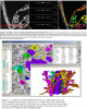
Data-analysis strategies for image-based cell profiling | Software | Workflow | Workflow of data-analysis strategies for image-based cell profiling. |
05/03/2023 - 15:53 |
|
|
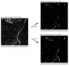
NET - Network Extraction Tool | Software | Collection | The ultimate goal of the NET framework is to make images of networks processable by computers. Therefore we want to have a pixel based image as input, as output we want a representation of the network visible in the image that retains as much information about the original network as possible. NET achives this by first segmenting the image and then vectorizing the network and then extracting information. The information we extract is |
05/03/2023 - 11:37 |
|
ORION | Software | Collection | ORION: Online Reconstruction and functional Imaging Of Neurons: segmentation and tracing of neurons for reconstruction. |
05/03/2023 - 15:20 |