
Contents
| Image | Title | Category | Type | Description | Updated |
|---|---|---|---|---|---|
| |
Diadem challenge | Dataset | Comprises six data set collections from different animal species, brain regions, neuron types, and visualization methods. |
05/03/2023 - 12:26 | |
|
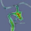
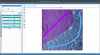
VMTK: Vascular Modeling Toolkit | Software | Collection | vmtk is a collection of libraries and tools for 3D reconstruction, geometric analysis, mesh generation and surface data analysis for image-based modeling of blood vessels. vmtk is composed of |
08/16/2018 - 17:07 |
| |
OpenCL | Software | Collection | OpenCL (Open Computing Language) is a framework for writing programs that execute across heterogeneous platforms consisting of central processing units (CPUs), graphics processing units (GPUs), digital signal processors (DSPs) |
01/28/2018 - 17:05 |

|
ClearGL | Software | Component | Facade API on top of JOGL (http://jogamp.org/jogl/www/) offering a simple interface for creating OpenGL contexts/windows, GLSL shader programs, and textures. Use it in your favourite JVM-based language. |
01/28/2018 - 13:16 |
| How to recover my password? | Forum topic | Look like there is no "password forgot" link in the login module. Would it be possible to add it? |
08/07/2018 - 13:15 | ||

|
CREMI challenge | Dataset | MICCAI Challenge on Circuit Reconstruction from Electron Microscopy Images |
01/28/2018 - 13:05 | |
| |
ClearVolume | Software | Component | ClearVolume is a real-time live 3D visualization library designed for high-end volumetric microscopes such as SPIM and DLSM microscopes. With ClearVolume you can see live on your screen the stacks acquired by your microscope instead of waiting for offline post-processing to give you an intuitive and comprehensive view on your data. The biologists can immediately decide whether a sample is worth imaging. ClearVolume can easily be integrated into existing Java, C/C++, Python, or LabVIEW based microscope software. |
01/28/2018 - 13:20 |

|
NeuroGPS-Tree | Software | Workflow | NeuroGPS-Tree is a workflow developed to reconstruct a neuronal population from a dense, large-scale data set. NeuroGPS-Tree is suitable for processing image stacks acquired by different image modalities. |
01/28/2018 - 19:29 |

|
Shiny - R package | Software | Collection | Shiny is an R package that makes it easy to build interactive web apps straight from R. |
01/28/2018 - 12:48 |

|
ClearCL | Software | Component | ClearCL is a Multi-backend Java Object Oriented Facade API for OpenCL. |
05/17/2023 - 15:35 |

|
MicrotubuleTracker in FIJI | Software | Collection, Component | MTrack is a tool, which detects, tracks, and measures the behavior of fluorescently labeled microtubules imaged by TIRF (total internal reflection fluorescence) microscopy. In such an in vitro reconstitution approach, stabilized, non-dynamic microtubule seeds serve as nucleation points for dynamically growing microtubules. |
05/03/2023 - 10:36 |
|
Trakem2 lens distortion correction | Software | Component | Calculates and corrects for lens-distortion models including chromatic abberation from confocal stacks. |
05/22/2018 - 02:35 |
| |
2D brain slice region annotation: SliceMap | Software | Workflow | SliceMap Whole brain tissue slices are commonly used in neurobiological research for analyzing pathological features in an anatomically defined manner. However, since many pathologies are expressed in specific regions of the brain, it is necessary to have an annotation of the regions in the brain slices. Such an annotation can be done by manual delineation, as done most often, or by an automated region annotation tool. |
05/02/2023 - 18:19 |

|
Globals for Images SimFCS 4 | Software | Collection | Software for analysis, visualization, simulation, and acquisition of data from spectroscopy and fluorescence microscopy. |
10/17/2018 - 18:47 |
| |
scenery | Software | Component | scenery is a scenegraphing and rendering library. It allows you to quickly create high-quality 3D visualisations based on mesh data. scenery contains both a OpenGL 4.1 and Vulkan renderer. |
05/17/2023 - 15:54 |
|
|
Multiview Reconstruction | Software | Collection | The Multiview Reconstruction software package enables users to register, fuse, deconvolve and view multiview microscopy images. The software is designed for lightsheet fluorescence microscopy (LSFM), but is applicable to any form of three or higher dimensional imaging modalities like confocal timeseries or multicolor stacks. |
01/30/2018 - 12:34 |

|
BigDataViewer | Software | Component | The BigDataViewer is a re-slicing browser for terabyte-sized multi-view image sequences. BigDataViewer was developed with multi-view light-sheet microscopy data in mind and integrates well with Fiji's SPIMage processing pipeline. |
01/30/2018 - 12:34 |

|
NeuriteTracer | Software | Workflow | "The plugin analyzes fluorescence microscopy images of neurites and nuclei of dissociated cultured neurons. Given user-defined thresholds, the plugin counts neuronal nuclei, and traces and measures neurite length."[...]" NeuriteTracer is a fast simple-to-use ImageJ plugin for the analysis of outgrowth in two-dimensional fluorescence microscopy images of neuronal cultures. The plugin performed well on images from three different types of neurons with distinct morphologies." |
10/28/2025 - 02:39 |
| |
intelligent Matrix Screener Remote Control | Software | Workflow | Integrates hardware control of Leica microscopes (via CAM), image analysis (e.g. via ImageJ, Matlab), and adaptive automatic screening of identified regions of interest. |
01/28/2018 - 12:16 |
|
SuRVoS | Software | Component | SuRVoS: Super-Region Volume Segmentation workbench A volume is first partitioned into Super-Regions (superpixels or supervoxels) and then interactively segmented by the user providing training annotations. SuRVoS can then learn from and extend the annotations to the whole volume. |
10/18/2018 - 17:38 |