
Contents
| Image | Title | Category | Type | Description | Updated | ||||
|---|---|---|---|---|---|---|---|---|---|
|
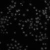
BBBC002v1 | Dataset | Five different samples of Drosophila melanogaster Kc167 cells were stained with Hoechst 33342, a DNA stain. The last sample (labeled nodsRNA) is of wild-type cells. Each of the other four samples (labeled 48, 340, Anillin, and mad2) has a different gene knocked down by RNAi. The sample preparation is described in more detail by [Carpenter et al. (Genome Biology, 2006)(http://genomebiology.biomedcentral.com/articles/10.1186/gb-2006-7-10-r1…) |
02/05/2019 - 10:24 | |||||

|
The NEMO dots assembly: Single-particle tracking and analysis | Software | Workflow | This workflow presents single-particle tracking in Fiji using Track-Mate, and track motility analysis in MATLAB using @msdanalyzer. |
05/24/2023 - 18:50 | ||||
| Documentation: Roles, Entry standard and how to curate (read first) | Basic page | Roles
|
05/15/2023 - 12:58 | ||||||
|
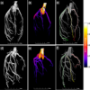
3D vessel segmentation of synchrotron phase contrast tomography | Software | Workflow | This workflow describes a semi-automatic image segmentation procedure for 3D reconstructions of the coronary arterial tree, after which how different morphometric features are automatically extracted, including vessel lumen diameter of the three main coronaries. |
05/03/2023 - 10:25 | ||||

|
CRImage | Software | Collection | CRImage a package to classify cells and calculate tumour cellularity CRImage provides functionality to process and analyze images, in particular to classify cells in biological images. Furthermore, in the context of tumor images, it provides functionality to calculate tumour cellularity. |
02/03/2019 - 19:41 | ||||

|
clij2 | Software | Collection | CLIJ2 is a GPU-accelerated image processing library for ImageJ/Fiji, Icy, Matlab and Java. |
04/29/2023 - 11:03 | ||||

|
Chromagnon | Software | Component | Image correction software for chromatic shifts in fluorescence microscopy |
10/27/2025 - 08:45 | ||||

|
Daybook2 | Software | Collection | Daybook 2 is the analysis software linked to argoligth slides. It tests the performance of microscopes on various levels: illumination homogeneity, field distortion, lateral resolving power, stage drift, chromatic aberrations, intensity response... It works with various file formats but requires the use of an argolight test slide. |
02/05/2019 - 12:17 | ||||
|
|
ConfocalCheck | Software | Component | Assess the performance of the lasers, the objective lenses and other key components required for optimum confocal operation. |
02/05/2019 - 12:16 | ||||

|
MIPs for PSFs | Software | Component | The macro generates orthogonal projections from bead images along the lateral and axial dimensions which are displayed using a customized look-up-table to color code intensities. A Gaussian curve is fit to the intensity profile of a fluorescent bead image and full-with-at-half-maximum (FWHM) values are extracted, and listed next to theoretical values for comparison. |
04/27/2023 - 16:53 | ||||

|
MetroloJ | Software | Collection | This plugin allows measuring relevant parameters which helps testing, following and comparing microscopes performances. This is achieved by extracting four indicators out of standardized images, acquired from standardized samples: the estimation of the detector sensitivity, the evaluation of the field illumination homogeneity, the system resolution, and finally the characterization of its spectral registration. |
05/03/2023 - 12:02 | ||||
|
|
MeVisLab XMarkerShortestPath module (Dijsktra shortest path) | Software | Component | 02/04/2019 - 18:00 | |||||
|
|
BoneJ | Software | Collection | 01/29/2019 - 21:59 | |||||
| |
MeVisLab | Software | Collection | 01/30/2019 - 11:02 | |||||
| video tutorial on 3D vessel segmentation of synchrotron phase contrast tomography | Training Material | In this tutorial video, a coronary arterial tree is used as the demo example to show in detail how the semi-automatic segmentation workflow, Carving from the open-source image analysis software ilastik, can be used. Tips on how and why a preprocessing is done, as well as parameter settings are provided. |
01/30/2019 - 11:05 | ||||||
|
Spimagine | Software | Component | Spimagine is a python package to interactively visualize and process time lapsed volumetric data as generated with modern light sheet microscopes (hence the Spim part). The package provides a generic 3D+t data viewer and makes use of GPU acceleration via OpenCL. If provides further an image processor interface for the GPU accelerated denoising and deconvolution methods of gputools. |
01/24/2019 - 17:15 | ||||
|
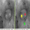
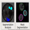
TEM ExosomeAnalyzer | Software | Workflow | TEM ExosomeAnalyzer is a program for automatic and semi-automatic detection of extracellular vesicles (EVs), such as exosomes, or similar objects in 2D images from transmission electron microscopy (TEM). The program detects the EVs, finds their boundaries, and reports information about their size and shape. The software has been developed in terms of project MUNI/M/1050/2013 and supported by Grant Agency of Masaryk University. |
01/21/2019 - 16:43 | ||||
|
|
Registrationshop | Software | Collection, Component | It is an interactive front-end visualization for registration software based on Elasix (VTK/ITK) |
01/21/2019 - 16:08 | ||||
|
SODA suite | Software | Component | Ensemble of blocks that implement SODA method for confocal and super-resolution microscopy, in 2 and 3 dimensions |
04/29/2023 - 15:51 | ||||
|
PartSeg | Software | Collection | There are many methods in bio-imaging that can be parametrized. This gives more flexibility |
01/16/2019 - 19:12 |