
Contents
| Image | Title | Category | Type | Description | Updated |
|---|---|---|---|---|---|
|
ThicknessTool | Software | Workflow | A plugin for the ImageJ platform that automates measurement of retinal nuclear layer thickness (e.g., outer nuclear layer (ONL), inner nuclear layer (INL)) by placing callipers perpendicular to the contour of segmented layers, enabling rapid, reproducible quantification across large images and multiple modalities. |
10/28/2025 - 02:05 |

|
BraiAn | Software | Collection, Workflow | BraiAn is an open-source suite of tools designed to simplify signal quantification, analysis and visualization of large datasets typically obtained in whole-brain imaging experiments, following registration to an atlas. The package consists of two separate modules. |
10/28/2025 - 06:45 |

|
Ultrack | Software | Collection, Workflow | Ultrack is a versatile and scalable cell tracking method designed to address the challenges of tracking cells across 2D, 3D, and multichannel timelapse recordings, especially in complex and crowded tissues where segmentation is often ambiguous. By evaluating multiple candidate segmentations and employing temporal consistency, Ultrack ensures robust performance under segmentation uncertainty. Ultrack's methodology is explained here. |
10/28/2025 - 01:44 |
|
SpotMAX | Software | Component | A generalist framework for multi-dimensional automatic spot detection and quantification. SpotMAX is designed to accomplish two tasks:
It supports 2D, 3D, 4D, and 5D data, i.e., z-stacks, timelapse, and multiple fluorescence channels (and combinations thereof). |
10/28/2025 - 01:53 |
|
Galaxy YOLO image segmentation | Software | Workflow | This workflow is the integration of YOLO (You Only Look Once) machine learning models, image pre-processing scripts and labeling tools within the Galaxy platform. Galaxy is an open, web-based platform used primarily for data analysis in computational biology, but it also has applications in image processing and other fields. How the Galaxy YOLO image segmentation tool works |
10/28/2025 - 02:17 |
|
Cell-ACDC | Software | Collection | Description from Github page: |
10/27/2025 - 09:18 |

|
MetaXpress | Software | Collection | MetaXpress or in full name "MetaXpress® High-Content Image Acquisition and Analysis Software" is a commercially available closed source software for high-content analysis from Molecular Devices, LLC.. The program is a kind of visually guided workflow programming environment. There is a programming module called CME (custom module editor) which lets one setup integrated workflows for bioimage analysis with visual feedback. It is designed for high-throughput in connection with a included database which stores the experimental data. |
10/28/2025 - 02:36 |
| |
python-ultralytics | Software | Collection | You can also install ultralytics directly from the Ultralytics GitHub repository. This can be useful if you want the latest development version. Ensure you have the Git command-line tool installed, and then run: |
10/27/2025 - 08:48 |
|
|
ABBA | Software | Collection | Aligning Big Brains & Atlases (ABBA) is a set of software components which allows users to register images of thin serial biological tissue sections, cut in any orientation (coronal, sagittal or horizontal) to atlases, usually brain atlases. ABBA is available as a Fiji plugin for performing registration; a QuPath extension is also available and recommended. Typically, a set of serial sections is defined as a QuPath project, that is registered within Fiji. |
10/27/2025 - 10:13 |
|
|
aNMJ-Morph | Software | Workflow | ImageJ macro script to streamline the original NMJ-morph methodology doi:10.1098/rsob.160240 |
10/28/2025 - 06:57 |
| |
microSAM | Software | Component | Tools for segmentation and tracking in microscopy build on top of Segment Anything. Segment and track objects in microscopy images interactively with a few clicks. |
10/27/2025 - 02:39 |
|
|
Placeholder Component Example | Software | Component | This is a placeholder component created for demonstration purposes. It serves as an example of how to create a minimal software entry in BIII. This component represents a basic image processing algorithm that can be used as part of a bioimage analysis workflow. |
10/27/2025 - 02:29 |
| |
YOLO | Software | Component | Ultralytics creates cutting-edge, state-of-the-art (SOTA) YOLO models built on years of foundational research in computer vision and AI. Constantly updated for performance and flexibility, our models are fast, accurate, and easy to use. |
10/27/2025 - 09:01 |
|
SNT | Software | Collection | SNT is ImageJ’s framework for tracing, visualization, quantitative analyses and modeling of neuronal morphology. For tracing, SNT supports modern multidimensional microscopy data, semi-automated and automated routines, and options for editing traces. For data analysis, SNT features advanced visualization tools, access to all major morphology databases, and support for whole-brain circuitry data. |
10/27/2025 - 03:14 |
|
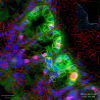
Movies of drosophila epithelia development, with manually annotated extrusions and divisions - From DeXtrusion publication | Dataset | Epithelia development movies. Drosophila pupal notum, marked with fluorescent E-cadherin |
08/21/2025 - 16:22 | |
|
|
Big-fish | Software | Component | Big-FISH is a python package for the analysis of smFISH images (2D/3D). It includes various methods to analyze microscopy images, such spot detection and segmentation of cells and nuclei. |
08/21/2025 - 14:42 |
|
Qupath Multiplex Analysis | Software | Workflow | Scripts that allow to automatize a multiplex analysis in qupath. The input is a multi channel image with one channel containing a staining of cell nuclei and the other channels containing markers for a specific molecule. The goal is to obtain a count of positive cells for the markers in the different channels and for the combinations of positive markers. The workflow also adds the total numbers of positive cells for each marker to the results table (cells positive for multiple markers are not counted positive for the individual markers in the original qupath result). |
01/08/2025 - 13:58 |
|
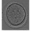
Segmentation of membrane of mouse, sea urchin and human oocytes from transmitted light images | Dataset | This dataset contains images acquired in transmitted light with different settings of mouse and human oocytes and sea urchin eggs, with the corresponding ground-truth of the membrane segmentation. Mouse oocyte images were taken before and during oocyte maturation (meiosis I). Some human oocyte images were taken during oocyte maturation (meiosis I), and some are M-II oocytes just after fertilization. Sea urchin images contains both fertilized and unfertilized eggs. |
11/13/2024 - 12:36 | |

|
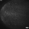
Movies of mouse oocyte maturation in transmitted light | Dataset | This dataset contains 466 movies of mouse oocytes maturation acquired in transmitted light every 3 min. Spatial resolution is 0.227 µm/pixel. |
11/13/2024 - 11:28 | |
| |
Oocytor | Software | Component | Fiji plugin to segment oocyte and zona pellucida contours from transmitted light images and extract hundreds of morphological features to describe numerically the oocyte. Segmentation is based on trained neural networks (U-Net) that were trained on both mouse and human oocytes (in prophase and meiosis I) acquired in different conditions. They are freely avaialable on the github repository and can be retrained if necessary. Oocytor also have options to extract hundreds of morphological/intensity features to characterize manually the oocyte (eg perimeter, texture...). |
11/13/2024 - 11:35 |